
Bonding to enamel no longer holds any secrets in orthodontics, but this is not the case for bonding to dentin. However, there are clinical situations where we apply the bonding protocol on the enamel, while we bond on the dentin. This can explain failures as in the following clinical case.
Bonding to enamel no longer holds any secrets in orthodontics, but this is not the case for bonding to dentin. However, there are clinical situations where we apply the bonding protocol on the enamel, while we bond on the dentin. This can explain failures as in the following clinical case.
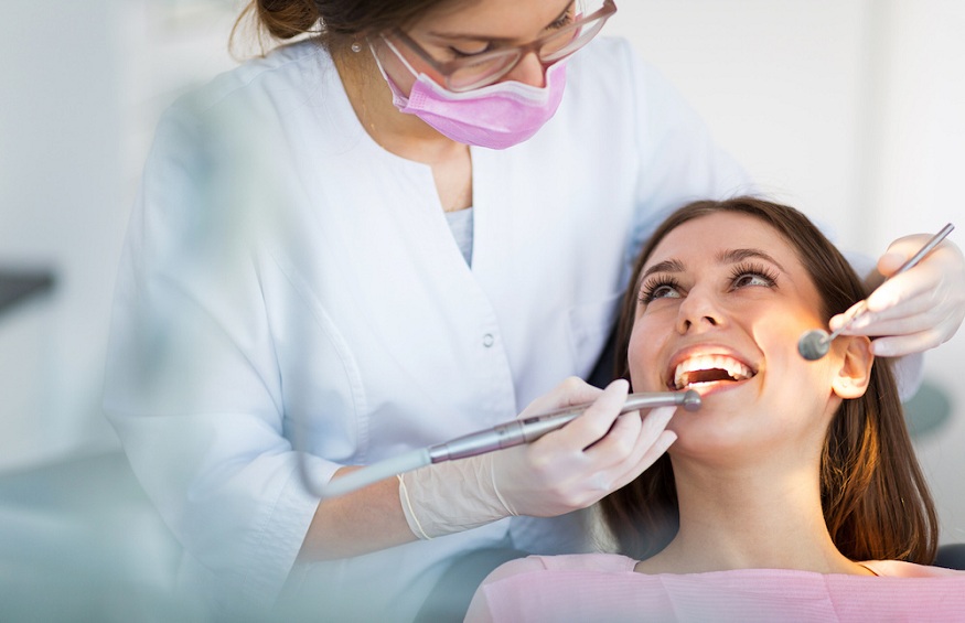
In the context of indirect lingual bonding, six anterior orthodontic locks remained in the transfer tray (fig. 1). The premolars and molars were however well bonded, although the protocol is identical. How can we explain this failure?
In a general context where the 45-year-old patient has dental sequelae from adolescent anorexia, we have to get used to carefully observing the dental surfaces on which we stick. Here we can note large dentinal areas. It is therefore a dentin bonding protocol that should have been applied in this context.
Here are four key points for optimizing adhesion to dentinal substrates in vestibular or lingual orthodontics.
Know how to distinguish eroded dentin in the active phase from eroded dentin in the inactive or sclerotic phase
The eroded dentin in the active phase, light yellow (fig. 2), is characterized by peritubular and intertubular demineralization. Thus, the surface exposes collagen fibers that are difficult for the adhesive to infiltrate [1].
The eroded dentin in the inactive phase is colored and sclerotic (fig. 3). It is a physiological response of the pulpo-dentine complex to aging and “slow” attacks. The defense mechanism of the pulpo-dentin complex induces a mineralized formation within the dentin, obliterating the tubules and forming a 14 µ thick hypermineralized layer of hydroxyapatite crystals on its surface [2].